Hacking the Life Sciences Classroom
This post is mirrored from The FabLearn Fellows Blog.
by Jaymes Dec
I recently helped to coordinate a “hackathon” with the Rockefeller University Science Outreach Program in part to address these questions:
- How can “making” or “STEAM” play out in the Life Sciences classroom?
- What role can digital design and fabrication tools have in the Life Sciences Classroom?
- How can physical computing tools and creative coding contribute to the Life Sciences classroom?
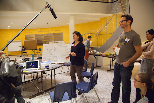
On a Saturday, we gathered about 30 educators, mostly life sciences teachers, but also technology and art teachers, from the NYC area. We had about 50% public teacher participation. We also invited 30 prominent biologists, biohackers, and engineers to spend the day tinkering, exploring, and prototyping project ideas with an abundance of materials and tools. Here is that list of supplies.
The morning started with a few short presentations in an auditorium. I spoke about Digital Design/Fabrication, and Physical/Creative Computing. My slides are here. Then we had Sebastian Kraves and Zeke Alvarez-Saavedra from miniPCR give a presentation about their project to make DNA amplification through the Polymerase Chain Reaction accessible to anyone. Finally, Sarah Weisberg from the BioBus, gave a presentation on their travelling Bio Lab and the MiScope, a portable digital microscope.
After the talks we had an “Ideas Fair”. In the weeks coming up to the event, we had gathered a few interesting DIY Bio ideas on this Pinterest page. These were printed on paper and hung around the room. During this portion of the day, several organizations and experts set up info booths around the room so that people could come and chat about hands-on life sciences activities. The miniPCR and BioBus people had spaces, as well as BioBuilder, The Makery, Genspace, and Harlem DNA Labs.
After about an hour of idea trading and pollination, everyone gathered for a group brainstorm session. Inspired by the format of the Constructing Modern Knowledge Summer Institute, we had people shout out ideas while runners wrote them on large post-it notes hung around the room.
There were many excellent ideas for projects. Here are a few: Hacking your own DIY Bio lab kit, a neuron model video game, natural selection simulation on Scratch, an attention measuring device, heart rate sensing, biofeedback device, 3D printing normal and mutated proteins, a Tamaguchi type game that simulates viruses, a birdhouse with sensors (tweeting birdhouse), and making music from proteins.
After a lunch break where we encouraged people to continue talking about the ideas, the rest of the day was dedicated to project time. Many people moved around the room from project to project until they sorted out which project they wanted to work on. As expected, some of the ideas got dropped, some new ideas formed, and some ideas organically combined. For the final hour, we moved from table to table as we did presentations and debriefs on each project. At the end we had a nice group conversation about some of the challenges around implementing these types of projects in the classroom.
The “highlight” projects were:
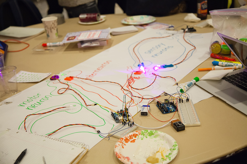
A model of different types of neurons using arduinos, leds, and photocells.
This team wanted to make a model that demonstrated how different types of neurons communicate across a system. They drew a network of large neurons on paper. Then they used LEDs to model the electrochemical signals getting passed across synapses. On the dendrites of each neuron, there were photocells that “received” the signals from the LEDs. Some neurons would fire only when they received signals from two other neurons. Some were inhibitor neurons that would prevent others from firing if they were activated. It was a fairly complex system. This group of about eight researchers and teachers had never worked with physical computing tools before! They split up into smaller groups and each group contributed by making a different neuron. It was really fun to watch them as they worked to calibrate threshold values and map analog input values to PWM values. They really seemed to enjoy the trial and error process.

Timelapse GIF of Daphnia pooping.
Sarah Weisberg from the BioBus, brought a MiScope, a portable digital microscope. A team of educators and scientists spent the afternoon watching Daphnia water fleas under the microscope. They dyed some algae and fed it to the Daphnia. Then they watched and took video as the food travelled all the way through the digestive system. They created a great animated GIF of the Daphnia pooping.
Measuring attention with biosensors and Scratch.
A group of teachers spent a few hours experimenting with different techniques to measure attention. They hacked a few off the shelf devices and used several different sensors to collect their data using Scratch. Stephen Lewis brought a few of his new MakeSense! boards. They allow you to easily get analog sensor data into Scratch. The group wrote a Scratch program that graphed data in real time. They collected data on heart rate and galvanic skin response.
Physiological response to eating hot peppers.
In that same group, one of the teachers was inspired by his students and their obsession with hot peppers and rankings on the Scoville Scale. He wanted to try to use sensors to measure the human bodies response to eating hot peppers. After a quick trip to the local market, another Fab Learn Fellow(!), Tracy Rudzitis volunteered to be the test subject. We could clearly see an increase in her heart rate as the capsaicin kicked in.
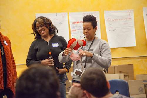
Sewed and knitted organs.
An arts educator from Brooklyn International High School coordinated a team to knit and sew a few human organs at actual scale. They made models of parts of the digestive system including the stomach, bladder, and intestine.
Prototype of Experimental device for studying circadian rhythms in crickets.
This team wanted to make a box with sound sensors that could collect data on the chirping rhythms of crickets. The idea was to create an artificial environment that made it easy to change single variables (vibrations, light cycle, sounds, etc) in the cage and study the effects on the circadian cycles in the insects. This was an ambitious project for the time allowed, but they did demonstrate their idea with a diorama including 3D printed crickets!
**
Too often the life sciences is taught through memorization of vocabulary words and labeling diagrams. These direct teaching methods suck the wonder and life out of the Life Sciences themselves!
It’s clear that “making” or “STEAM” can play a significant part in breathing life back into the Life Sciences classroom. Creating real working models of biological systems and collecting and analysing actual biodata is true science, treats students like real citizen scientists, and exposes them to the joys and mysteries of the life sciences.
One classic method of teaching life sciences is to have students build models of biological systems. While this is certainly a better method than memorizing vocabulary or labelling diagrams, these models are too often static and don’t represent the dynamic nature of these systems. As we were planning this event, I often talked about going “beyond the model”. But it turns out that physical and creative computing tools allow learners to quickly prototype dynamic models of biological systems that are so much more instructive than static models. It’s one thing to explore the interaction of different types of neurons through a drawing. But when you have to make a working model of the way they communicate and inhibit communication with each other, you really do understand the system at a deeper and more appreciative level.
A common concern that we hear from teachers, especially in the life sciences, is that they don’t have enough time to “cover the content” and do hands-on activities as well. I think that this view misses the true point of teaching and learning. School should not be about cramming in as much information as possible so that you can “cover” your content. School should be able creating opportunities for students to uncover or discover content so that they can construct their own knowledge. This is precisely what making and hands-on activities do so well. I can imagine that sewing or knitting bodily organs would take a lot of time. But if some of the reasons for learning about the systems of the body include thinking about and appreciating the purposes and complexity of these incredible systems, perhaps slowing down and exploring the scale, textures, and connections between organs is not a bad idea. Teachers have to become more comfortable with sacrificing content for discovery and exploration.
Overall this event was a great success. We explored and addressed the posed questions about technology, making, and the life sciences. Additionally, the event served as further proof that this format of bringing together experts and educators to tinker and experiment with creative uses of technology works really well. In the near future, we hope to organize more events like this one. I think it would be interesting to target groups of teachers from disparate subjects like: History and Science, or Music and Math.